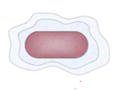
STRUCTURE OF EUBACTERIAL CELL
In
eukaryotic cells, there is extensive compartmentalization of cytoplasm
through the presence of membrane-bound organelles. Eukaryotic cells
possess an organized nucleus with a nuclear envelope. In addition,
eukaryotic cells have a variety of complex locomotory and cytoskeletal
structures. Their genetic material is organized into chromosomes
A eukaryote is any cellular or organism that possesses a surely described nucleus. The eukaryotic cell has a nuclear membrane that surrounds the nucleus, in which the well-defined chromosomes (bodies containing the hereditary material) are located. Eukaryotic cells additionally include organelles, together with mitochondria (mobile strength exchangers), a piece of Golgi equipment (secretory tool), an endoplasmic reticulum (a canal-like organelle of membranes within the cell), and lysosomes (digestive apparatus within many cell types).
There are numerous exceptions to this, but; for example, the absence of mitochondria and a nucleus in red blood cells and the shortage of mitochondria
The bacterial structure is very simple but they are very complex in
behavior.
Cell Envelope
The cell envelope consists of a tightly bound three-layered
structure.
(1) Outermost glycocalyx
(2) Cell wall
(3) Cell membrane
Although each layer of the envelope performs a distinct
function they act together as a single protective unit
Glycocalyx
- Slime layer:- Thin, Loose, Rough Sticky Mainly polysaccharides
Function of Glycocalyx
- Protects the bacteria from w.b.c.(leukocytes)
- Helps in colony formation.
Cell Wall
Bateria cell wall is made up of peptidoglycan(Amino acid+polsaccharide).
L-form: Bacterial cell wall can be removed by lysozyme enzyme.
When the bacterial cell wall is removed artificially then these bacteria
are called L - forms (Lister form)
Cell membrane
Made up of lipoprotein like the eukaryotic membrane.
Cytoplasm
In bacterial cytoplasm membrane-bound cell organelles viz.
Mitochondria, Chloroplast E.R., Lysosome, Golgibody,
Microbodies, etc. are absent.
Mesosomes
Mesosome (also known as chondroid) is a
special membranous structure formed by the
extension or infoldings or invaginations of the plasma membrane into the cell. These extensions are in the form of vesicles,
tubules, and lamellae. Analogous to mitochondria
(Oxidative enzymes are found in mesosomes)
Functions of Mesosomes :
1. They help in cell respiration, and increase the surface area of
the plasma membrane and enzymatic content.
2. Help in DNA replication and distribution to daughter cells
during cell division.
3. Cell wall material secretion or cell wall formation
4. Cell division
Storage granules/Inclusion bodies
Reserve material in a prokaryotic cell.
Not bound by any membrane system and lie free in the
cytoplasm.
A. Glycogen granules: They store carbohydrate
B. Volutin granules: These are also known as metachromatic
granules. The volutin granules are phosphate
polymers and function as a storage reservoir for phosphate.
Chromatin material (Nucleoid/Genophore)
DNA is double-stranded, circular, and naked.
Plasmid
Plasmids can replicate independently.
Plasmids are of many types based on their function or
phenotypic character.
(i) F-plasmid (fertility factor): Based on the presence or absence of the 'F' plasmid, there are two types of bacteria.
(a) F
+ Cells, carrying 'F' plasmid, act as donors and are called F
+
or
male.
(b) F
– Cells, lacking 'F' plasmid, act as recipients and are called F
–
or
female.
(ii) R-factor: Resistant to certain antibiotics.
When the 'F" plasmid is attached to the main DNA, it is designated
as Episome and this type of cell is known as Hfr (High-frequency recombinant) cell.
First of all H.C. Gram differentiated bacteria based on staining
Gram-positive
- The bacteria remain purple-colored with Gram staining
even after washing with alcohol.
- The cell wall is single-layered.
- The cell wall of peptidoglycan is 20–80 nm.
- Murein (Peptidoglycan)
70–80%.
- The wall is smooth
- Basal body of the flagellum
2 rings (S & M).
- Mesosomes are quite
prominent
- Few pathogenic bacteria
belong to the Gram-positive group.
- Teichoic acid present in cell wall
Gram-negative
- The bacteria do not retain the stain when washed with alcohol.
- The cell wall is bilayered.
- The cell wall of peptidoglycan 8–12 nm
- Murein (Peptidoglycan) 10–20%
- Wall is wavy and in contact with the cell membrane at a few loci.
- Basal body of the flagellum 4 rings (L, P, S & M)
- Mesosomes are less prominent.
- Most of the pathogenic bacteria belong to the Gram-negative group.
- Teichoic acid was absent.


0 Comments
Post a Comment
If you have any doubt, please let me Know